
Memory training: White matters
Throughout life the human brain is constantly changing and modifying itself to adapt to the changing demands of the environment. This so-called cognitive plasticity is an inherent characteristic of the brain that essentially reflects one’s ability to learn. Older individuals tend to vary substantially to the degree that they show improvement through cognitive intervention and therefore document varying degrees of cognitive plasticity. Indeed, older individuals tend to be characterised by great variability in terms of how much they may benefit from mental training.
This underscores the need to understand both the brain mechanisms that predispose older individuals to benefit greatly from mental training, as well as the brain mechanisms underlying the inability to benefit from such training.
In our latest research paper we conceptualised cognitive plasticity as the extent to which older individuals benefited from a memory training intervention.
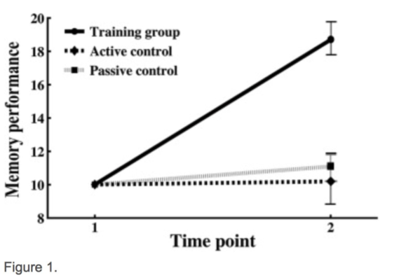
For some years our group has been running an extensive training study (see NCP project) where our participants have undergone either intensive memory training to remember word lists, or been assigned to a control group that received no specific memory training. During memory training participants learnt to use a technique known as the Method of Loci, whereby in order to memorize a list of objects in sequential order one must visualise each of the objects and mentally place them along a familiar route. By this method, each object to-be-remembered becomes associated with a particular location along a journey, and this enables easier recall.
Participants underwent MRI scans at the onset of the intervention, and every ten weeks during the study.
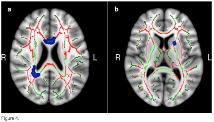
As predicted, only the group that underwent memory training had shown an improvement in memory score by the end of the study period (above figure), demonstrating our memory-training paradigm to be effective. In the brain’s white matter tracts it was found that relative to older adults, younger adults tended to show a greater degree of white matter integrity (as inferred by diffusion measurements of molecular motion) across many regions of the brain. In addition, the analysis led to the identification of 3 specific areas of white-matter that related to the degree of memory improvement from the training we administered (below figure).
This was the key finding, as it indicates that individuals with a lager degree of white matter microstructural integrity as measured before the training period, benefitted more from the training and thus were able to improve their memory scores to a greater extent.Therefore the integrity of the brain’s white matter tracts could be a potential marker for one’s ability to adapt in response to cognitive demands during ageing.
Citation
98. White-matter integrity as a marker for cognitive plasticity in ageing ![]()
Glasø de Lange, A; Bråthen, A. C; Grydeland, H; Sexton, C; Johansen-Berg, H; Andersson, J; … Walhovd, K. (2016)
Getting connected
The two hemispheres of the brain are connected by an extensive bundle of nerve fibers known as the corpus callosum. This enables neural information both to be exchanged and shared between the two halves of the brain, or possibly delegated to a greater extent to one hemisphere more than the other. If the corpus callosum is impaired in any way then there will be a concurrent lack of inter-hemispheric communication and a large mental deficit. Therefore corpus collosal development represents a crucial step in healthy brain maturation in children; the structure typically shows a growth spurt during the first 2-3 years of life, achieving adult-like levels of function by middle to late childhood.
However, all previous research examining the structural development of the corpus callosum in middle to late childhood has also included adolescent children and even young adults. Therefore the results of these previous growth studies will have been swayed by the inclusion of older children, and the statistics are likely to be more representative of older children given their often greater number. In a recent paper LCBC investigated corpus callosal growth for the first time using a sample exclusively composed of children in middle to late childhood (aged 4-11 years), using brain scans from 428 children (approx. 50% gender split) with follow-up scans from 304.
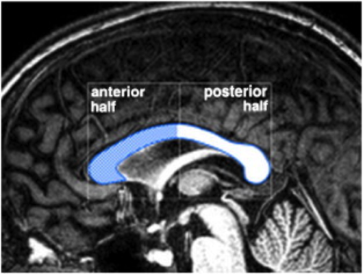
The team found that the corpus callosum of children shows significant increases in thickness relative to overall brain size (i.e. after taking into account the fact that overall brain size increases during development) between 4-11 years. The growth rate found amounted to a 0.19mm growth in thickness per year in posterior regions (see top image) where the age effect was strongest.
This higher growth rate of the posterior part of the corpus callosum agrees with development research in general; it is known that the more posterior parts of the brain (visual and sensory areas) mature faster than anterior regions, and the posterior half of the corpus callosum links posterior brain regions. It therefore makes sense that we observed a more pronounced growth spurt in posterior corpus callosum, simply because brain changes associated with maturing visual and sensory systems are among the first to occur in young children. Thus this region-specific thickening of the nerve fibers connecting these systems is likely an effect of maturing sensory brain areas.
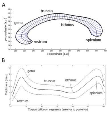
Top figure from Kompus, Kalpouzos & Westerhausen (2011). Brain Research. doi:10.1016/j.brainres.2011.08.052
Citation
97. Selective increase in posterior corpus callosum thickness between the age of 4 and 11 years ![]()
Cerebral Cortex
Westerhausen, R; Fjell, A. M; Krogsrud, S. K; Rohani, D; Skranes, J; Håberg, A & Walhovd, K. (2016).
Staying connected
Advanced mental functions rely on large-scale networks of communicating brain cells, and the coordination of information from widespread regions of the brain. It has been proposed that the communication-efficiency of these networks is something that declines as we age, and that this may help to explain phenomena such as slowing reaction times with advanced age, as well as degradation of so-called higher-order mental functions.
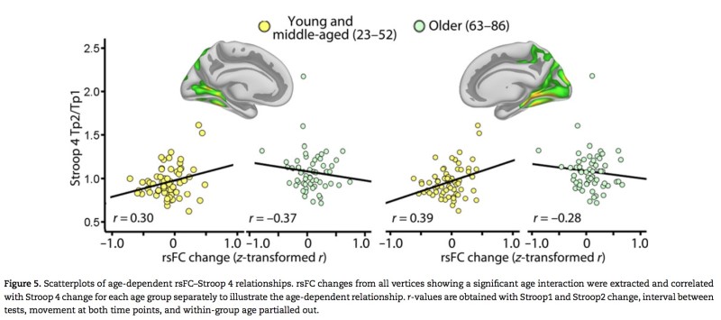
To test the hypothesis of a more ‘disconnected brain’ with greater age, we followed 119 young, middle-aged and older (63+) participants with repeated brain scans and took measurements of brain connectivity between cortical grey-matter regions while at rest (functional connectivity), as well as measurements of white-matter tract integrity (structural connectivity) — the major information highways in the brain composed of bundles of myelinated axons that allow far-flung cortical regions to connect. We also tested participants on a well-known attentional control task. This task is known to heavily recruit frontal brain regions that are more associated with higher-order mental abilities; patients with damage to such brain regions tend to perform poorly on this task.
We found that executive higher-order cognitive functions showed a clear decline with age over time, and this was to a far greater extent than other, more basic mental functions. MRI-derived measures of brain structural and functional connectivity were both found to relate to this reduction in executive functioning between the two test time-points, and 82.5% of the age-related decline observed in our study could be explained by connectivity changes over time.
As a caveat it should be noted that all aging research is inherently flawed because it involves quantifying changes from one time to another whilst allowing the influence of time to take it’s natural course. Therefore, since age and time cannot be subjected to experimental manipulation, our results are correlational in nature and therefore do not allow us to draw causal conclusions. Nevertheless, we believe our results may support the view that as we age, the human brain becomes more ‘disconnected’, and this appears to happen in concert with a decline in higher-order mental functioning.
Citation
The disconnected brain and executive function decline in aging ![]()
Cerebral Cortex
Fjell, A. M; Sneve, M. H; Grydeland, H; Storsve, A. B. & Walhovd, K. B. (2016).
What determines brain plasticity?

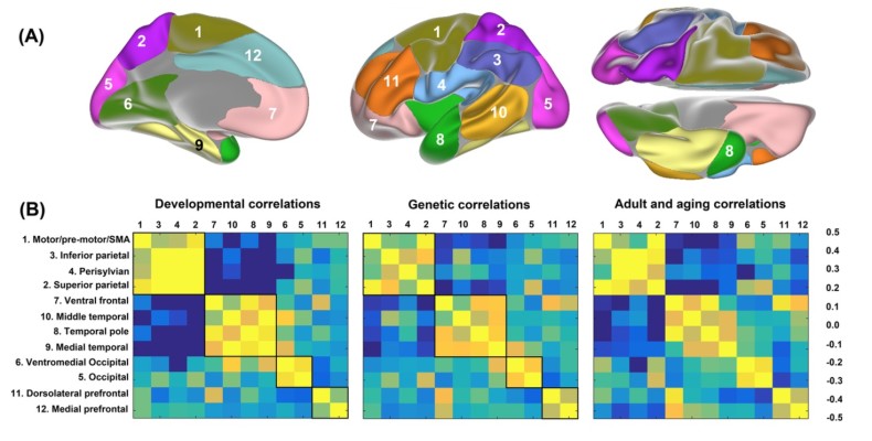
What is it that determines the brain’s potential for flexibility? In a new perspective paper, LCBC examines the case for brain plasticity — the potential for long-lasting change in the way the brain both functions and is structured. Brain plasticity is the mechanism that allows us to learn new skills and information, and to adapt our behavior to the ever-changing demands of the world. The paper explores the possible pre-requisites for whether a brain region can demonstrate high or low levels of plasticity. In particular, it is discussed how brain regions exhibiting low levels of expansion throughout human evolutionary history and low levels of genetic heritability may also be characterized by higher levels of plasticity. In addition, brain areas showing greater variability of structural change across individuals throughout the lifespan, and those characterized by higher degrees of inter-individual myelin content may also show higher potential for plastic change. The medial temporal lobe is one such critical area that satisfies these conditions.
Citation
Walhovd, K. B., Westerhausen, R., de Lange, A. M. G., Bråthen, A. C. S., Grydeland, H., Engvig, A., & Fjell, A. M. (2015). Premises of plasticity—And the loneliness of the medial temporal lobe. NeuroImage.
Genetic influence on lifespan brain development – new PNAS paper
Our group has just published a new research paper in the prestigious journal Proceedings of the National Academy of Sciences of the United States of America. The study investigates how genetically determined neurodevelopmental events can have consequences for the structural architecture of the brain that span the duration of human life, and thus how both neurodevelopment and aging relate to the underlying genetic organization of the cerebral cortex. A total of 1633 brain scans were used from a longitudinal sample of 974 participants aged between 4 and ~89 years of age, in addition to 773 scans of children below the age of 12. A separate sample of adult twins was used to obtain brain maps indicating the regions of the cerebral cortex that showed the highest degree of genetic overlap in terms of cortical thickness. Cortical changes as a result of both early brain maturation and aging were found to adhere closely to the genetic organization of the cerebral cortex. This finding suggests that predetermined genetic factors contribute to cortical changes occurring throughout life.
In the media
Study results suggest genetics play a role in later life cerebral cortex thickness (MedicalXpress)
Citation
Fjell, A. M., Grydeland, H., Krogsrud, S. K., Amlien, I., Rohani, D. A., Ferschmann, L., … & Walhovd, K. B. (2015). Development and aging of cortical thickness correspond to genetic organization patterns. Proceedings of the National Academy of Sciences, 201508831.