Tag Archive for: New paper

What determines brain plasticity?

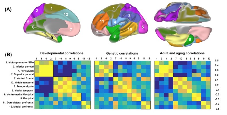
What is it that determines the brain’s potential for flexibility? In a new perspective paper, LCBC examines the case for brain plasticity — the potential for long-lasting change in the way the brain both functions and is structured. Brain plasticity is the mechanism that allows us to learn new skills and information, and to adapt our behavior to the ever-changing demands of the world. The paper explores the possible pre-requisites for whether a brain region can demonstrate high or low levels of plasticity. In particular, it is discussed how brain regions exhibiting low levels of expansion throughout human evolutionary history and low levels of genetic heritability may also be characterized by higher levels of plasticity. In addition, brain areas showing greater variability of structural change across individuals throughout the lifespan, and those characterized by higher degrees of inter-individual myelin content may also show higher potential for plastic change. The medial temporal lobe is one such critical area that satisfies these conditions.


Citation

Walhovd, K. B., Westerhausen, R., de Lange, A. M. G., Bråthen, A. C. S., Grydeland, H., Engvig, A., & Fjell, A. M. (2015). Premises of plasticity—And the loneliness of the medial temporal lobe. NeuroImage.

Genetic influence on lifespan brain development – new PNAS paper

Dev_and_genetic_org1

Our group has just published a new research paper in the prestigious journal Proceedings of the National Academy of Sciences of the United States of America. The study investigates how genetically determined neurodevelopmental events can have consequences for the structural architecture of the brain that span the duration of human life, and thus how both neurodevelopment and aging relate to the underlying genetic organization of the cerebral cortex. A total of 1633 brain scans were used from a longitudinal sample of 974 participants aged between 4 and ~89 years of age, in addition to 773 scans of children below the age of 12. A separate sample of adult twins was used to obtain brain maps indicating the regions of the cerebral cortex that showed the highest degree of genetic overlap in terms of cortical thickness. Cortical changes as a result of both early brain maturation and aging were found to adhere closely to the genetic organization of the cerebral cortex. This finding suggests that predetermined genetic factors contribute to cortical changes occurring throughout life.


In the media

Study results suggest genetics play a role in later life cerebral cortex thickness (MedicalXpress)


Citation

Fjell, A. M., Grydeland, H., Krogsrud, S. K., Amlien, I., Rohani, D. A., Ferschmann, L., … & Walhovd, K. B. (2015). Development and aging of cortical thickness correspond to genetic organization patterns. Proceedings of the National Academy of Sciences, 201508831.

Development of white matter tracts in children

krogsrud_2015

LCBC has just published a new research paper in the journal NeuroImage. The paper uses diffusion tensor imaging – a measure of the extent of diffusion of water molecules across the brain which links with the extent of neuronal myelination – to investigate the development of the brain’s white matter tracts in children aged between 4 and 11 years across two scan intervals. Linear patterns of white matter tract development were observed globally across the brain between scan intervals, with greater changes observed in later-maturing frontal brain regions.


Citation

Krogsrud, S. K., Fjell, A. M., Tamnes, C. K., Grydeland, H., Mork, L., Due-Tønnessen, P., … & Walhovd, K. B. (2015). Changes in white matter microstructure in the developing brain-a longitudinal diffusion tensor imaging study of children from 4 to 11 years of age. NeuroImage.

Go to publications page


Memory, aging, and the brain at rest

Screen Shot 2015-09-23 at 14.47.41

LCBC has just published a new research paper in the journal Neurobiology of Aging. The study addresses the question of whether levels of functional connectivity between diverse brain regions in the resting brain can help to explain differences in episodic memory ability with age. Using a longitudinal follow-up design (after 3.5 years) across many ages, the difference in memory score between baseline and 3.5 years was found to be related to resting state functional connectivity measures – an index of brain connectivity at rest. It was thus found that changes in episodic memory ability over time related to connectivity levels in the resting brain, and that this also varied in an age-dependent manner.


Citation

Fjell, A. M., Sneve, M. H., Grydeland, H., Storsve, A. B., de Lange, A. M. G., Amlien, I. K., … & Walhovd, K. B. (2015). Functional connectivity change across multiple cortical networks relates to episodic memory changes in aging. Neurobiology of Aging.

Go to publications page


Development of children born to mothers with mental health problems: subcortical volumes and cognitive performance at 4½ years

European Child and Adolescent Psychiatry; pdf

Bjørnebekk, A; Siqveland, T; Haabrekke, K J; Moe, V; Slinning, K; Fjell, A M & Walhovd, K B (2014)

Poor sleep quality links with cortical thinning

White matter reveals the brain’s age

The cerebral cortex thins with age

The self-regulating developing brain